In-house 3D printing is increasingly being used in dental clinics and laboratories as intra-oral scanners, CAD software, 3D printers, and printable resins for prostheses resins and aligners have advanced and become more accessible for routine clinical use. Current applications include 3D models, surgical guides, craniomandibular orthopaedic positioning appliances, crowns, bridges, dentures, aligners, retainers, mock-ups, implant-supported provisional restorations and implant superstructures.
At our clinic, in-house production has been part of daily practice for more than five years, and 3D printing now plays an important role in all aspects of our implant treatment. In edentulous and nearly edentulous cases, including all-on-4, all-on-5 and all-on-6 treatments, the use of 3D-printed provisional restorations is valuable for assessing and refining occlusion, jaw position and aesthetics. 3D printing offers design flexibility, precision, reproducibility and shorter turnaround times and can also help reduce laboratory costs. Staining and glazing can further enhance the appearance of printed restorations. This article outlines several 3D-printing workflows used in our clinical practice.
Fabrication of 3D models
STL files generated from intra-oral scans can be imported into 3D-printing software and used to produce dental models. In orthodontic workflows, orthodontic analysis software can be used to design a treatment timeline, generate the relevant STL files and 3D-print models for each step. In conventional thermoforming workflows, aligner sheets can then be thermoformed over these printed models to fabricate aligners (Fig. 1).
Fabrication of craniomandibular orthopaedic positioning appliances
The target jaw position at which centric relation coincides with centric occlusion is first determined. The intra-oral scan data can then be imported into orthodontic analysis software and used to design a craniomandibular orthopaedic positioning appliance. The design is then exported to a 3D printer. In patients with craniomandibular disorders, this type of appliance is used to achieve muscular deprogramming (Fig. 2).
Fabrication of surgical guides
The guide workflow differs between dentate and edentulous cases. In both, intra-oral scan data is combined with CT or CBCT DICOM data in implant planning software. The resulting surgical guide design can then be exported and 3D-printed (Fig. 3).
Fig. 3: 3D-printed surgical guide with metal sleeves shown intra-orally.
Fabrication of definitive crowns
Crown designs created in dedicated CAD software can be 3D-printed using a crown resin. A wide range of shades is available, and fabrication can be performed using either a single-layer or a double-layer technique. To enhance aesthetics, staining and glazing processes are applied.
In recent years, not only have 3D printers advanced, but significant progress has also been made in prosthetic resins. These improvements include increased ceramic content for enhanced aesthetics, greater load-bearing capacity and improved tensile strength. When selecting materials for clinical use, it is essential to consider whether they have sufficient mechanical strength and fracture resistance under occlusal loading and whether they can reproduce the colour and light-transmitting properties of natural teeth. For example, Tera Harz C & B R-191 (Graphy), a resin developed used for definitive crowns and provisional restorations, has been designed to meet these requirements and is therefore suitable for in-house 3D printing (Figs. 4a–c).
Fig. 4a: Crowns 3D-printed from Tera Harz C & B R-191. Crowns on the build platform after printing.
Fig. 4b: Prepared maxillary central incisors.
Fig. 4c: Crowns in situ.
Fabrication of provisional restorations
Implant position data is captured either digitally, using scan bodies and an intra-oral scanner, or conventionally, by creating stone models from silicone impressions. This data is then combined with occlusal vertical dimension records obtained from chairside provisional implant restorations, which are fabricated intra-orally using an auto-polymerising acrylic resin. The combined datasets are then used to design provisional restorations in CAD software and produce them using a 3D printer. After printing, staining and glazing can be used to improve the aesthetics of the provisional restorations. A key advantage of this workflow is the relatively short fabrication time, which makes it easier to refine occlusion, the occlusal plane, tooth morphology and tooth shade during the provisional phase.
In my experience, Tera Harz C & B R-191 has shown favourable clinical performance in the fabrication of provisional restorations. It combines durability with high aesthetic quality, making it well suited for provisional use under functional load (Figs. 5a–f).
Fig. 5a: Implant-supported provisional restorations 3D-printed from Tera Harz C & B R-191. Restorations on the build platform after printing.
Fig. 5f: All-on-5 provisional restoration shown after intra-oral placement.
Fabrication of Shape Memory Aligners
Facial photographs, intra-oral scan data, and CT or CBCT data are imported into orthodontic analysis software for diagnosis and tooth movement simulation. The resulting treatment set-up is uploaded to aligner design software, where the aligners are prepared for 3D printing and then sent to a 3D printer for fabrication (Figs. 6a–c).
Fig. 6a: Shape Memory Aligners 3D-printed from Tera Harz Clear TA-28 on the build platform.
Fig. 6b: Aligner after post-processing.
Fig. 6c: Aligners shown intra-orally.
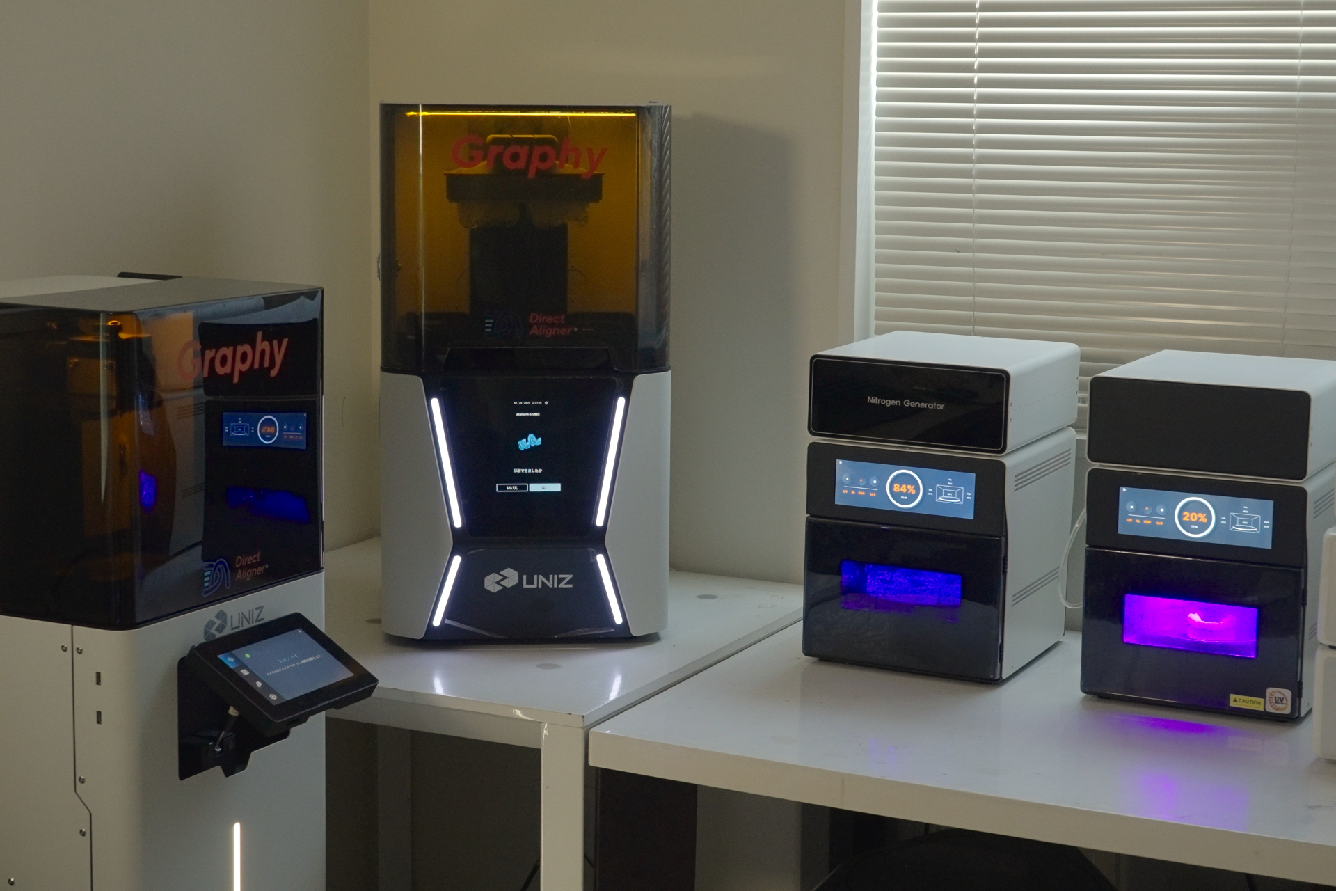
Fig. 7: UBEE 3D printer and Tera Harz Cure post-polymerisation unit used in the Shape Memory Aligner workflow.
Compared with conventional thermoforming methods, this approach allows greater customisation. In the in-house workflow described here, Shape Memory Aligners (Tera Harz Clear TA-28) were printed using a UBEE 3D printer (UNIZ Technology), and post-polymerisation was performed under nitrogen in a Tera Harz Cure unit (Fig. 7).
Tera Harz Clear TA-28 has been designed to exhibit shape memory behaviour at an intra-oral temperature of approximately 37 °C. It delivers light, continuous force for approximately two weeks, enabling efficient tooth movement. Because the aligner can extend over the cervical undercut areas, it provides excellent retention and enhances the efficiency of tooth movement. I have used these aligners without attachments in selected cases. In addition, the resin softens with heat, making insertion and removal easier for patients.
Conclusion
3D printing is a central component of the shift towards in-house dental production and will continue to develop as a dynamic field. It enables the creation of 3D models, surgical guides, craniomandibular orthopaedic positioning appliances, crowns, implant-supported provisional restorations and aligners with high precision and efficient workflows. For clinics, the main benefits include shorter turnaround times and reduced reliance on external laboratory production.
The digitisation of dentistry has unlocked many potential applications in modern clinical and technical practice. 3D printing is becoming an increasingly ...
HONG KONG, China: CyberSmile, a new training centre for computer-assisted implant surgery, opened in Hong Kong earlier this year. Established by Drs James ...
In today’s rapidly evolving dental landscape, 3D printing is emerging as a game-changer, transforming the way dental professionals approach patient care. ...
As a certified dental technician, I have worked in, managed and owned several laboratories around the world. This has exposed me to many different analogue,...
BANGKOK, Thailand: Held on 27 and 28 March in the Thai capital, the Osstem World Meeting 2026 Bangkok highlighted the latest clinical strategies and ...
HONG KONG: Researchers from the University of Hong Kong have developed a personalised 3D-printed mouth guard that they say has plaque removal results ...
Maxillary reconstruction has entered a new era. What was once mainly a matter of closing a defect or fitting an obturator has now become a carefully ...
Evaluating how implant depth, crown design, angulation, and patient factors influence implant outcomes in a 5-year follow-up study at SEGi University.
...
Complete edentulism is an irreversible condition that is a known marker of disease burden for oral health.1 Edentulism significantly influences both oral ...
Education
Live webinar Thu. 15 October 2026 10:00 am EST (New York)



 International / International
 Brazil / Brasil
 Canada / Canada
 Latin America / Latinoamérica
 USA / USA
 Austria / Österreich
 Bosnia and Herzegovina / Босна и Херцеговина
 Bulgaria / България
 Croatia / Hrvatska
 Czech Republic & Slovakia / Česká republika & Slovensko
 France / France
 Germany / Deutschland
 Greece / ΕΛΛΑΔΑ
 Hungary / Hungary
 Italy / Italia
 Netherlands / Nederland
 Nordic / Nordic
 Poland / Polska
 Portugal / Portugal
 Romania & Moldova / România & Moldova
 Slovenia / Slovenija
 Serbia & Montenegro / Србија и Црна Гора
 Spain / España
 Switzerland / Schweiz
 Turkey / Türkiye
 UK & Ireland / UK & Ireland
 China / 中国
 India / भारत गणराज्य
 Pakistan / Pākistān
 Vietnam / Việt Nam
 Israel / מְדִינַת יִשְׂרָאֵל
 Algeria, Morocco & Tunisia / الجزائر والمغرب وتونس
 Middle East / Middle East

 Dr. Sinan GhishanRegister now1CELive webinar
 Dr. Sonia PresenciaLive webinar
 Dr. John Tsourounakis DDS, Cert. Perio, MS, FRCD(C)Register now1CELive webinar

 Dr. Varkha Rattu, Cat EdneyRegister now1CE











To post a reply please login or register